- Home /
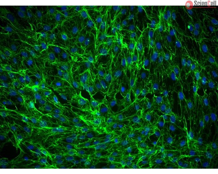
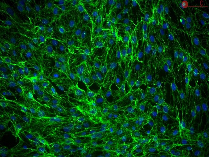
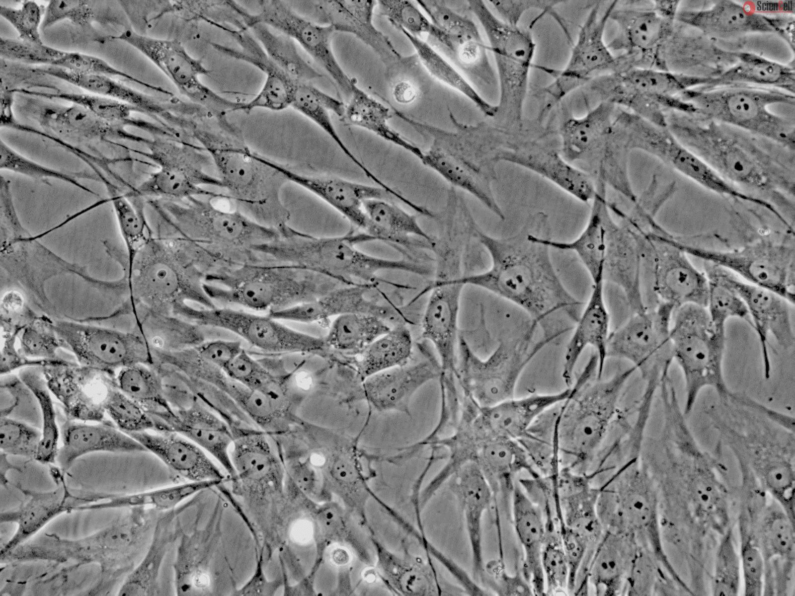
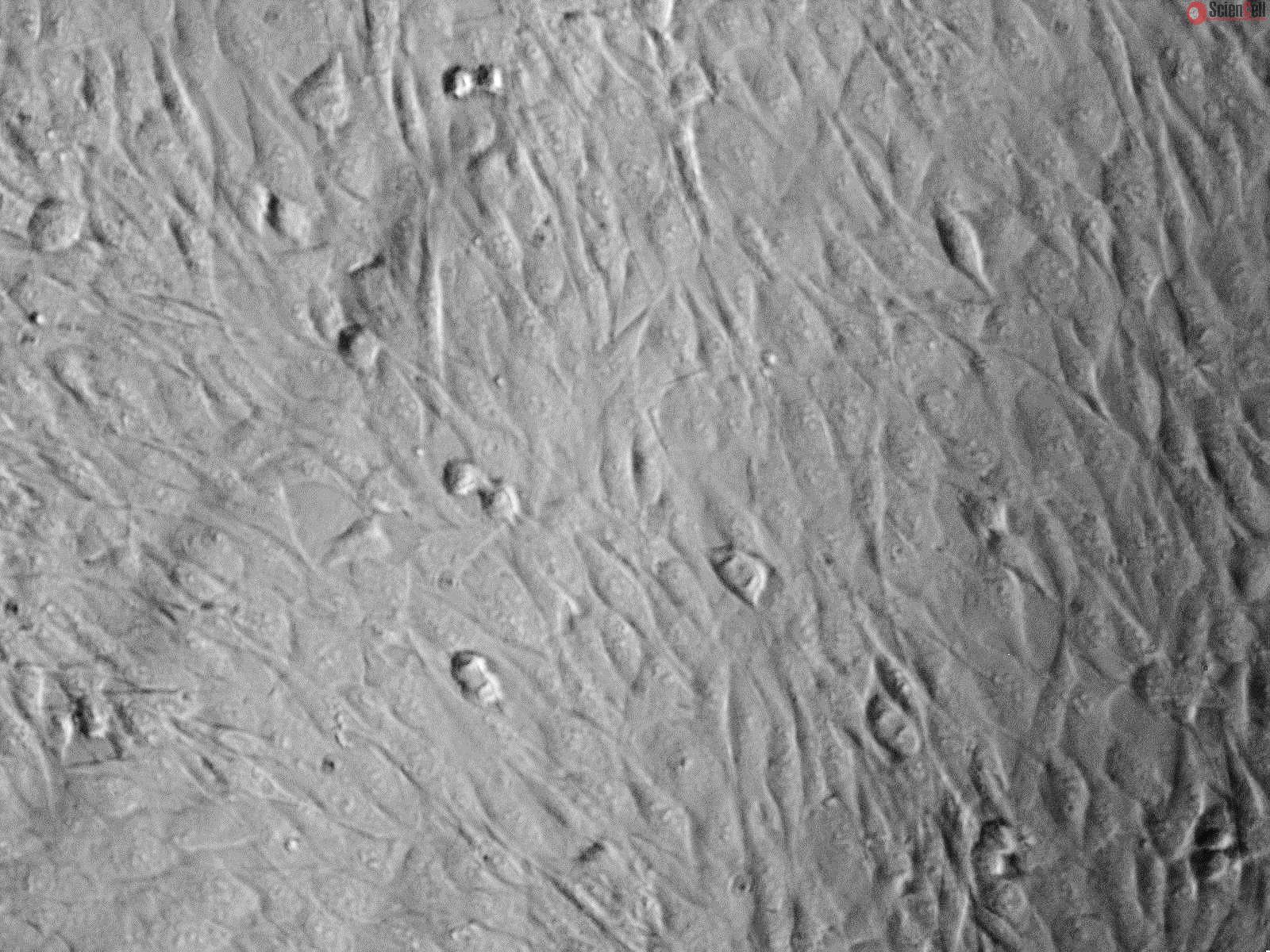
- Human Dermal Fibroblasts-Fetal-Mitomycin C treated
Fibroblasts are mesenchymal cells derived from the embryonic mesoderm. They have been extensively used for a wide range of cellular and molecular studies as they are one of easiest types of cells to grow in culture. Their durability also makes them amenable to a variety of manipulations ranging from studies employing gene transfection to microinjection. In general, fibroblasts secrete a non-rigid extracellular matrix which is rich in type I and/or type III collagen. There is evidence showing that fibroblasts in various organs are intrinsically different. Dermal fibroblasts switch from a proliferative, migratory phase to a contractile, matrixremodeling phase during wound healing. In addition, they secrete large quantities of hyaluronan in response to inflammatory stimuli.
HDF-f-mt from ScienCell Research Laboratories are isolated from fetal human skin. HDF-f-mt are cryopreserved at passage one and delivered frozen. Each vial contains > 5 x 10^5 cells in 1 ml volume. HDF-f-mt are characterized by their spindle morphology and immunofluorescence with antibody specific to fibronectin. HDF-f-mt are negative for HIV-1, HBV, HCV, mycoplasma, bacteria, yeast, and fungi. HDF-f-mt are guaranteed to further culture under the conditions provided by ScienCell Research Laboratories; however, HDF-f-mt are not recommended for expanding as mitomycin C treatment prevents further cell proliferation.
Recommended Medium
It is recommended to use Fibroblast Medium (FM, Cat. #2301) for culturing HDF-f-mt in vitro.
Catalog No. | 2350 |
---|---|
Country of Manufacture | United States |
Product Code | HDF-f-mt |
Size/Quantity | 1 x 10^5 cells/vial |
Product Use | This product is for research use only. It is not approved for use in humans, animals, or in vitro diagnostic procedures. |
Storage | Directly and immediately transfer cells from dry ice to liquid nitrogen upon receiving and keep the cells in liquid nitrogen until cell culture needed for experiments. |
Shipping Info | Dry ice. |
References | [1] Conrad, G. W., Hart, G. W., Chen, Y. (1977) Differences in vitro between fibroblast-like cells from cornea,
heart, and skin of embryonic chicks. J. Cell Sci. 26:119-137. [2] Gabbiani, G., Rungger-Brandle, E., The fibroblast. In Tissue Repair and Regeneration (L. E. Glynn, ed.), pp 1- 50. Handbook of Inflammation, Vol. 3. Amsterdam, Elsevier, 1981. [3] Stair S, Carlson KW, Shuster S, Wei ET, Stern R (2002) Mystixin peptides reduce hyaluronan deposition and edema formation. Eur J Pharmacol 30;450(3):291-6. |
No Publication available at this time
CAT. NO. | CODE | DESCRIPTION | PRICE | Qty | |
---|---|---|---|---|---|
REAGENTS | |||||
0913 | FibroF | FibroFectagen |
$253.00
|
||
MOLECULAR BIOLOGY | |||||
2354 | HDF-f-mt cDNA | Human Dermal Fibroblast-fetal-mitomycin C treated cDNA |
$411.00
|
||
2356 | HDF-f-mt Lysate | Human Dermal Fibroblast-fetal-mitomycin C treated Lysate |
$398.00
|
||
2359 | HDF-f-mt gDNA | Human Dermal Fibroblast-fetal-mitomycin C treated Genomic DNA |
$398.00
|