We have previously reported that the majority of phospholipase A2 (PLA2) activity in rabbit ventricular myocytes is membrane-associated, calcium-independent (iPLA2), sele... More
We have previously reported that the majority of phospholipase A2 (PLA2) activity in rabbit ventricular myocytes is membrane-associated, calcium-independent (iPLA2), selective for arachidonylated plasmalogen phospholipids and inhibited by the iPLA2-selective inhibitor bromoenol lactone (BEL). Here, we identified the presence of iPLA2 in rabbit ventricular myocytes, determined the full length sequences for rabbit iPLA2beta and iPLA2gamma and compared their homology to the human isoforms. Rabbit iPLA2beta encoded a protein with a predicated molecular mass of 74 kDa that is 91% identical to the human iPLA2beta short isoform. Full length iPLA2gamma protein has a predicated molecular mass of 88 kDa and is 88% identical to the human isoform. Immunoblot analysis of iPLA2beta and gamma in membrane and cytosolic fractions from rabbit and human cardiac myocytes demonstrated a similar pattern of distribution with both isoforms present in the membrane fraction, but no detectable protein in the cytosol. Membrane-associated iPLA2 activity was inhibited preferentially by the R enantiomer of bromoenol lactone [(R)-BEL], indicating that the majority of activity is due to iPLA2gamma. Less
Information from the brain travels back and forth along peripheral nerves in the form of electrical impulses generated by neurons and these impulses have repetitive patte... More
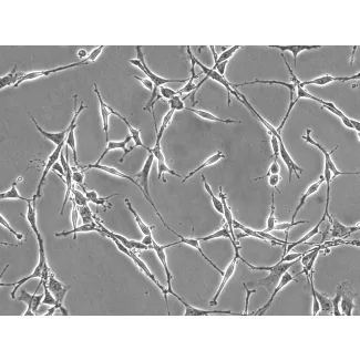
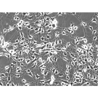
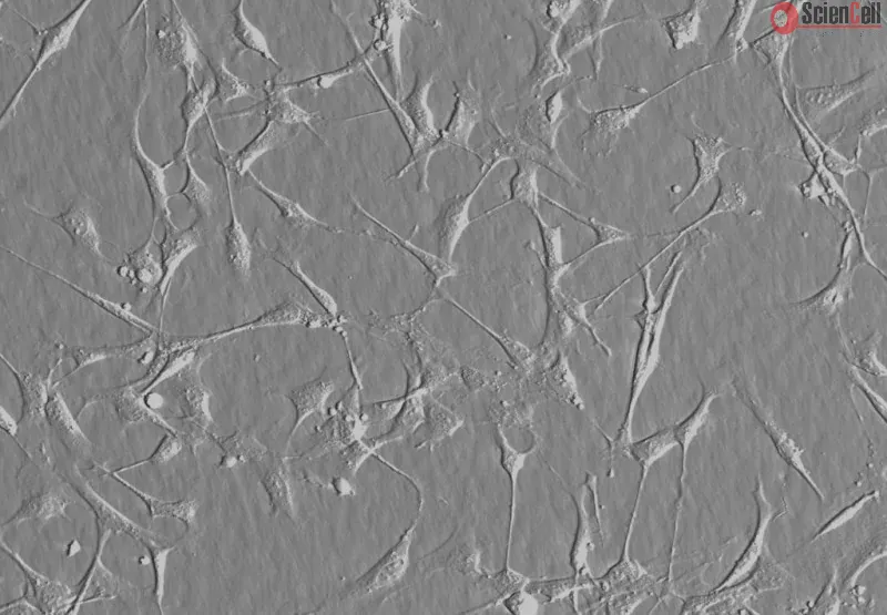
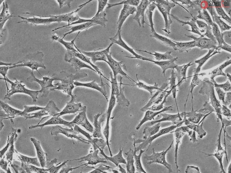
Information from the brain travels back and forth along peripheral nerves in the form of electrical impulses generated by neurons and these impulses have repetitive patterns. Schwann cells in peripheral nerves receive molecular signals from axons to coordinate the process of myelination. There is evidence, however, that non-molecular signals play an important role in myelination in the form of patterned electrical impulses generated by neuronal activity. The role of patterned electrical impulses has been investigated in the literature using co-cultures of neurons and myelinating cells. The co-culturing method, however, prevents the uncoupling of the direct effect of patterned electrical impulses on myelinating cells from the indirect effect mediated by neurons. To uncouple these effects and focus on the direct response of Schwann cells, we developed an in vitro model where an electroconductive carbon fiber acts as an artificial axon. The fiber provides only the biophysical characteristics of an axon but does not contribute any molecular signaling. In our "suspended wire model", the carbon fiber is suspended in a liquid media supported by a 3D printed scaffold. Patterned electrical impulses are generated by an Arduino 101 microcontroller. In this study, we describe the technology needed to set-up and eventually replicate this model. We also report on our initial in vitro tests where we were able to document the adherence and ensheath of human Schwann cells to the carbon fiber in the presence of patterned electrical impulses (hSCs were purchased from ScienCell Research Laboratories, Carlsbad, CA, USA; ScienCell fulfills the ethic requirements, including donor's consent). This technology will likely make feasible to investigate the response of Schwann cells to patterned electrical impulses in the future. Less
Background. Chronic inflammatory demyelinating polyneuropathy (CIDP) is often associated with chronic disability, which can be accounted to incomplete regeneration of inj... More
Background. Chronic inflammatory demyelinating polyneuropathy (CIDP) is often associated with chronic disability, which can be accounted to incomplete regeneration of injured axons. We hypothesized that Schwann cell support for regenerating axons may be altered in CIDP, which may account for the poor clinical recovery seen in many patients. Less
Vestibular schwannoma (VS) is a benign, slow-growing cranial tumor that originates from the hypertrophy of Schwann cells. The majority of sporadic VS are unilateral, and ... More
Vestibular schwannoma (VS) is a benign, slow-growing cranial tumor that originates from the hypertrophy of Schwann cells. The majority of sporadic VS are unilateral, and the mechanisms underlying VS tumorigenesis are not fully understood. The human neurofibromin 2 (NF2) gene encodes the tumor suppressor protein merlin and the NF2 transcript can be alternatively spliced to form numerous isoforms. The present study investigated human Schwann cells (HSCs) at the mRNA and protein level to understand the function of the alternative splicing (AS) isoform of NF2. The total RNA of HSCs was isolated and the full-length coding sequence of NF2 was amplified. The amplified products were excised from agarose gels, purified and sequenced. NF2 at a protein level was assayed by immunoprecipitation and western blot analysis. The full-length and spliced NF2 forms were amplified by polymerase chain reaction (PCR) from the HSC complementary DNA and ligated into eukaryotic expression vector pcDNA3.1(+). The plasmids were transfected into the HSC HEI-193 cell line and cell proliferation assays were performed using Cell Counting Kit-8. PCR analysis using HSC total RNA as a template revealed the presence of a shortened NF2 transcript, which was due to splicing at the 3′-end of the NF2 mRNA. Sequence analysis confirmed that this AS isoform omitted exons 11, 12, 13, 14, 15 and 16. Immunoprecipitation and western blot analysis demonstrated that the AS isoform was highly expressed in the HSCs at 38 kDa, while the wild-type (WT) isoform, which was expected at 66 kDa, was undetectable. Transfection and cell proliferation assays revealed that the WT isoform exhibited significant growth inhibition, while the AS isoform did not suppress cell growth. In conclusion, the present study detected AS NF2 isoforms in HSC for the first time, and investigated the function of the principle AS isoform. The present study suggests that although HSCs have an undetectable level of WT isoform of the NF2 protein merlin, they are not merlin-null, since they express the AS isoform. Although the AS merlin isoform has no suppressive effect on cell growth, certain mechanisms may exist that underlie this phenomenon, and this may be associated with the genesis and development of VS. Keywords: vestibular schwannoma, human Schwann cells, NF2, alternative splicing Less
Background Radical prostatectomy (RP) carries the risk of erectile dysfunction (ED) due to cavernous nerve (CN) injury. Schwann cells are essential for the maintenance of... More
Background Radical prostatectomy (RP) carries the risk of erectile dysfunction (ED) due to cavernous nerve (CN) injury. Schwann cells are essential for the maintenance of integrity and function of peripheral nerves such as the CNs. We hypothesize that brain-derived neurotrophic factor (BDNF) activates the Janus kinase (JAK)/(signal transducer and activator of transcription) STAT pathway in Schwann cells, not in neuronal axonal fibers, with the resultant secretion of cytokines from Schwann cells to facilitate nerve recovery. Methods Using four different cell lines—human neuroblastoma BE(2)-C and SH-SY5Y, human Schwann cell (HSC), and rat Schwann cell (RSC) RT4-D6P2T—we assessed the effect of BDNF application on the activation of the JAK/STAT pathway. We also assessed the time response of JAK/STAT pathway activation in RSCs and HSCs after BDNF treatment. We then assayed cytokine release from HSCs as a response to BDNF treatment using oncostatin M and IL6 as markers. Results We showed extensive phosphorylation of STAT3/STAT1 by BDNF at high dose (100 pM) in RSCs, with no JAK/STAT pathway activation in human neuroblastoma cell lines. The time response of JAK/STAT pathway activation in RSCs and HSCs after BDNF treatment showed an initial peak at shortly after treatment and then a second higher peak at 24–48 hours. Cytokine release from HSCs increased progressively after BDNF application, reaching statistical significance for IL6. Conclusions We demonstrated for the first time the indirect mechanism of BDNF enhancement of nerve regeneration through the activation of JAK/STAT pathway in Schwann cells, rather than directly on neurons. As a result of BDNF application, Schwann cells produce cytokines that promote nerve regeneration. Keywords: Brain-derived neurotrophic factor (BDNF), Janus kinase/signal transducer and activator of transcription (JAK/STAT), Schwann cells, nerve regeneration, cavernous nerve injury (CN injury) Less
Neural vascular insufficiency plays an important role in diabetic peripheral neuropathy (DPN). Peroxisome proliferative-activated receptor (PPAR)α has an endothelial pro... More
Neural vascular insufficiency plays an important role in diabetic peripheral neuropathy (DPN). Peroxisome proliferative-activated receptor (PPAR)α has an endothelial protective effect related to activation of PPARγ coactivator (PGC)-1α and vascular endothelial growth factor (VEGF), but its role in DPN is unknown. We investigated whether fenofibrate would improve DPN associated with endothelial survival through AMPK-PGC-1α-eNOS pathway. Fenofibrate was given to db/db mice in combination with anti-flt-1 hexamer and anti-flk-1 heptamer (VEGFR inhibition) for 12 weeks. The db/db mice displayed sensory-motor impairment, nerve fibrosis and inflammation, increased apoptotic cells, disorganized myelin with axonal shrinkage and degeneration, fewer unmyelinated fibers, and endoneural vascular rarefaction in the sciatic nerve compared to db/m mice. These findings were exacerbated with VEGFR inhibition in db/db mice. Increased apoptotic cell death and endothelial dysfunction via inactivation of the PPARα-AMPK-PGC-1α pathway and their downstream PI3K-Akt-eNOS-NO pathway were noted in db/db mice, human umbilical vein endothelial cells (HUVECs) and human Schwann cells (HSCs) in high-glucose media. The effects were more prominent in response to VEGFR inhibition. In contrast, fenofibrate treatment ameliorated neural and endothelial damage by activating the PPARα-AMPK-PGC-1α-eNOS pathway in db/db mice, HUVECs and HSCs. Fenofibrate could be a promising therapy to prevent DPN by protecting endothelial cells through VEGF-independent activation of the PPARα-AMPK-PGC-1α-eNOS-NO pathway. Less
Chemotaxis signals between hepatic stellate cells (HSC) and sinusoidal endothelial cells (SEC) maintain hepatic vascular homeostasis and integrity and also regulate chang... More
Chemotaxis signals between hepatic stellate cells (HSC) and sinusoidal endothelial cells (SEC) maintain hepatic vascular homeostasis and integrity and also regulate changes in sinusoidal structure in response to liver injury. Our prior studies have demonstrated that the bidirectional chemotactic signaling molecules EphrinB2 and EphB4 are expressed in HSC. The aim of our present study was to explore whether and how the EphrinB2/EphB4 system in HSC could promote SEC recruitment, which is essential for sinusoidal structure and remodeling. Stimulation of human HSC (hHSC) with chimeric agonists (2 microg/ml) of either EphrinB2 or EphB4 (EphrinB2 Fc or EphB4 Fc, respectively) significantly increased VEGF mRNA levels in hHSC as assessed by quantitative PCR, with respective small interfering RNAs for EphrinB2 and EphB4 inhibiting this increase (P < 0.05, n = 3). EphrinB2 agonist-induced increase in VEGF mRNA levels in hHSC was associated with increased phosphorylation of Erk and was significantly blocked by U0126 (20 microM), an inhibitor of MEK, which is a kinase upstream from Erk (P < 0.05, n = 3). The EphB4 agonist also significantly increased human VEGF promoter activity (P < 0.05, n = 3) as assessed by promoter reporter luciferase assay in transfected LX2-HSC. This was associated with upregulation of the vasculoprotective transcription factor, Kruppel-like factor 2 (KLF2). In Boyden chamber assays, conditioned media from hHSC stimulated with agonists of EphrinB2 or EphB4 increased SEC chemotaxis in a VEGF-dependent manner, compared with control groups that included basal media with agonists of EphrinB2, EphB4, or HSC-conditioned media from HSC in absence of agonist stimulation (P < 0.05, n = 3). EphB4 expression was detected in situ within liver sinusoidal vessels of rats after carbon tetrachloride-induced liver injury. In summary, activation of the EphrinB2/EphB4 signaling pathway in HSC promotes chemotaxis of SEC through a pathway that involves Erk, KLF2, and VEGF. These studies identify EphrinB2 or EphB4 as a key intermediary that links HSC signal transduction pathways with angiogenesis and sinusoidal remodeling. Less
Background: Increased mitogen-activated protein kinase (MAPK) phosphorylation has been detected in peripheral nerve of human subjects and animal models with diabetes as w... More
Background: Increased mitogen-activated protein kinase (MAPK) phosphorylation has been detected in peripheral nerve of human subjects and animal models with diabetes as well as high-glucose exposed human Schwann cells, and have been implicated in diabetic peripheral neuropathy. In our recent studies, leukocytetype 12/15-lipoxygenase inhibition or gene deficiency alleviated large and small nerve fiber dysfunction, but not intraepidermal nerve fiber loss in streptozotocin-diabetic mice. Methods: To address a mechanism we evaluated the potential for pharmacological 12/15-lipoxygenase inhibition to counteract excessive MAPK phosphorylation in mouse and cell culture models of diabetic neuropathy. C57Bl6/J mice were made diabetic with streptozotocin and maintained with or without the 12/15-lipoxygenase inhibitor cinnamyl-3,4-dihydroxy-α-cyanocinnamate (CDC). Human Schwann cells were cultured in 5.5 mM or 30 mM glucose with or without CDC. Results: 12(S) HETE concentrations (ELISA), as well as 12/15-lipoxygenase expression and p38 MAPK, ERK, and SAPK/JNK phosphorylation (all by Western blot analysis) were increased in the peripheral nerve and spinal cord of diabetic mice as well as in high glucose-exposed human Schwann cells. CDC counteracted diabetes-induced increase in 12(S)HETE concentrations (a measure of 12/15-lipoxygenase activity), but not 12/15-lipoxygenase overexpression, in sciatic nerve and spinal cord. The inhibitor blunted excessive p38 MAPK and ERK, but not SAPK/ JNK, phosphorylation in sciatic nerve and high glucose exposed human Schwann cells, but did not affect MAPK, ERK, and SAPK/JNK phosphorylation in spinal cord. Conclusion: 12/15-lipoxygenase inhibition counteracts diabetes related MAPK phosphorylation in mouse and cell culture models of diabetic neuropathy and implies that 12/15-lipoxygenase inhibitors may be an effective treatment for diabetic peripheral neuropathy. Keywords: Diabetes; Lipoxygenase; Mitogen-Activated Protein Kinase; Neuropathy; Schwann Cells. Less
Background: Lyme neuroborreliosis (LNB), caused by the spirochete Borrelia burgdorferi, affects both the peripheral and the central nervous systems. Radiculitis or nerve ... More
Background: Lyme neuroborreliosis (LNB), caused by the spirochete Borrelia burgdorferi, affects both the peripheral and the central nervous systems. Radiculitis or nerve root inflammation, which can cause pain, sensory loss, and weakness, is the most common manifestation of peripheral LNB in humans. We previously reported that rhesus monkeys infected with B. burgdorferi develop radiculitis as well as inflammation in the dorsal root ganglia (DRG), with elevated levels of neuronal and satellite glial cell apoptosis in the DRG. We hypothesized that B. burgdorferi induces inflammatory mediators in glial and neuronal cells and that this inflammatory milieu precipitates glial and neuronal apoptosis. Methods: To model peripheral neuropathy in LNB we incubated normal rhesus DRG tissue explants with live B. burgdorferi ex vivo and identified immune mediators, producer cells, and verified the presence of B. burgdorferi in tissue sections by immunofluorescence staining and confocal microscopy. We also set up primary cultures of DRG cells from normal adult rhesus macaques and incubated the cultures with live B. burgdorferi. Culture supernatants were subjected to multiplex ELISA to detect immune mediators, while the cells were evaluated for apoptosis by the in situ TUNEL assay. A role for inflammation in mediating apoptosis was assessed by evaluating the above phenomena in the presence and absence of various concentrations of the anti-inflammatory drug dexamethasone. As Schwann cells ensheath the dorsal roots of the DRG, we evaluated the potential of live B. burgdorferi to induce inflammatory mediators in human Schwann cell (HSC) cultures. Results: Rhesus DRG tissue explants exposed to live B. burgdorferi showed localization of CCL2 and IL-6 in sensory neurons, satellite glial cells and Schwann cells while IL-8 was seen in satellite glial cells and Schwann cells. Live B. burgdorferi induced elevated levels of IL-6, IL-8 and CCL2 in HSC and DRG cultures and apoptosis of sensory neurons. Dexamethasone reduced the levels of immune mediators and neuronal apoptosis in a dose dependent manner. Conclusion: In this model, B. burgdorferi induced an inflammatory response and neuronal apoptosis of DRG. These pathophysiological processes could contribute to peripheral neuropathy in LNB. Less
Herein, we performed microarray experiments in Schwann cells infected with live M. leprae and identified novel differentially expressed genes (DEG) in M. leprae infected ... More
Herein, we performed microarray experiments in Schwann cells infected with live M. leprae and identified novel differentially expressed genes (DEG) in M. leprae infected cells. Also, we selected candidate genes associated or implicated with leprosy in genetic studies and biological experiments. Forty-seven genes were selected for validation in two independent types of samples by multiplex qPCR. First, an in vitro model using THP-1 cells was infected with live Mycobacterium leprae and M. bovis bacillus Calmette-Guérin (BCG). In a second situation, mRNA obtained from nerve biopsies from patients with leprosy or other peripheral neuropathies was tested. We detected DEGs that discriminate M. bovis BCG from M. leprae infection. Specific signatures of susceptible responses after M. leprae infection when compared to BCG lead to repression of genes, including CCL2, CCL3, IL8 and SOD2. The same 47-gene set was screened in nerve biopsies, which corroborated the down-regulation of CCL2 and CCL3 in leprosy, but also evidenced the down-regulation of genes involved in mitochondrial metabolism, and the up-regulation of genes involved in lipid metabolism and ubiquitination. Finally, a gene expression signature from DEG was identified in patients confirmed of having leprosy. A classification tree was able to ascertain 80% of the cases as leprosy or non-leprous peripheral neuropathy based on the expression of only LDLR and CCL4. A general immune and mitochondrial hypo-responsive state occurs in response to M. leprae infection. Also, the most important genes and pathways have been highlighted providing new tools for early diagnosis and treatment of leprosy. Less
Cell-based therapy has achieved promising functional recovery for peripheral nerve repair. Although Schwann cells (SCs) and bone marrow derived mesenchymal stromal cells ... More
Cell-based therapy has achieved promising functional recovery for peripheral nerve repair. Although Schwann cells (SCs) and bone marrow derived mesenchymal stromal cells (BM-MSCs) are the main cell source for nerve tissue engineering, the clinical application is limited because of donor site morbidity, the invasive procedure, and the decreased number of SCs and BM-MSCs. Wharton's jelly-derived mesenchymal stem cells (WJMSCs) could be a promising cell source for nerve tissue engineering because they are easily accessible and their use has no ethical issues. We investigated the phenotypic, molecular and functional characteristics of WJMSCs differentiated along a Schwann-cell lineage. Cultured WJMSCs were isolated from human umbilical cord, and the undifferentiated WJMSCs were confirmed by the detection of MSC-specific cell-surface markers. WJMSCs treated with a mixture of glial growth factors (basic fibroblast growth factor, platelet-derived growth factor and forskolin) adopted a spindle-like morphology similar to SCs. Immunocytochemical staining, RT-PCR analysis, and Western blot analysis revealed that the treated cells expressed the glial markers glial fibrillary acidic protein, p75, S100 and P0 and indicative of differentiation. On co-culture with dorsal root ganglia neurons, the differentiated WJMSCs enhanced the number of sprouting neurites and neurite length in dorsal root ganglia neurons. Furthermore, using enzyme-linked immunosorbent assay and RT-PCR methodology, we found differentiated WJMSCs secrete and express neurotrophic factors, including brain-derived neurotrophic factor (BDNF), nerve growth factor (NGF), and neurotrophin-3 (NT-3). Quantification of neurite outgrowth from PC12 cells grown in differentiated WJMSCs-conditioned media demonstrates that the neurite length is significantly more than control medium and undifferentiated WJMSCs group. WJMSCs can be differentiated into cells that are Schwann-like in terms of morphologic features, phenotype, and function and could be suitable Schwann-cell substitutes for nerve repair in clinical applications. Copyright © 2010 Elsevier Inc. All rights reserved. Less
Varicella-zoster virus (VZV) and herpes simplex virus (HSV) are prevalent neurotropic herpesviruses that cause various nervous system diseases. Similar to other enveloped... More
Varicella-zoster virus (VZV) and herpes simplex virus (HSV) are prevalent neurotropic herpesviruses that cause various nervous system diseases. Similar to other enveloped viruses, membrane fusion is an essential process for viral entry. Therefore, identification of host molecules that mediate membrane fusion is important to understand the mechanism of viral infection. Here, we demonstrate that myelin-associated glycoprotein (MAG), mainly distributed in neural tissues, associates with VZV glycoprotein B (gB) and promotes cell-cell fusion when coexpressed with VZV gB and gH/gL. VZV preferentially infected MAG-transfected oligodendroglial cells. MAG also associated with HSV-1 gB and enhanced HSV-1 infection of promyelocytes. These findings suggested that MAG is involved in VZV and HSV infection of neural tissues. Keywords: herpes simplex virus, neurotropism, membrane fusion, Varicella-zoster virus, virus entry Less
Previous studies have demonstrated that merlin acts as a tumor suppressor by blocking Ras-mediated signaling. However, the mechanism by which merlin controls cell prolife... More
Previous studies have demonstrated that merlin acts as a tumor suppressor by blocking Ras-mediated signaling. However, the mechanism by which merlin controls cell proliferation has remained obscure. Here we show that merlin deficient tumors exhibited loss of p21, concomitant with elevated CDKs/cyclin D1 levels in sporadic vestibular schwannomas (VS) from clinic patients. Likewise, silencing of merlin gene expression in the cell lines resulted in down-regulation of p21. Furthermore, we find that merlin-enhanced p21 protein stability, rather than increased RNA accumulation, was responsible for the elevated p21 levels. Interestingly, p21 was required to maintain merlin levels and the inhibitory effect of merlin on Ras signaling was partially overridden by knockdown of p21. Consistent with the observation that over-expression of merlin arrested cell growth at G1-phase, the current study indicates that merlin exerts its antiproliferative effect, at least in part, by maintaining p21 expression, and loss of p21 is a prominent feature of merlin deficient schwannomas. Copyright 2010 IBRO. Published by Elsevier Ltd. All rights reserved. Less
This study evaluated the role of 12/15-lipoxygenase, which converts arachidonic acid to 12(S)- and 15(S)-hydroxyeicosatetraenoic acids, in nitrosative stress in the perip... More
This study evaluated the role of 12/15-lipoxygenase, which converts arachidonic acid to 12(S)- and 15(S)-hydroxyeicosatetraenoic acids, in nitrosative stress in the peripheral nervous system and peripheral prediabetic and diabetic neuropathies. The experiments were performed in C57BL6/J mice made diabetic with streptozotocin or fed a high-fat diet and in human Schwann cells cultured in 5.5 or 30 mM glucose. 12/15-Lipoxygenase overexpression and activation were present in sciatic nerve and spinal cord of diabetic and high-fat diet-fed mice, as well as in human Schwann cells cultured in high concentrations of D-, but not L-glucose. 12/15-Lipoxygenase inhibition by cinnamyl-3,4-dihydroxy-alpha-cyanocinnamate (8 mg kg(-1) day(-1) sc, for 4 weeks after 12 weeks without treatment) alleviated the accumulation of nitrated proteins in the sciatic nerve and spinal cord, and large and small nerve fiber dysfunction, but not intraepidermal nerve fiber loss. 12/15-Lipoxygenase gene deficiency alleviated nitrosative stress and nerve conduction deficit, but not small sensory fiber neuropathy, in high-fat diet-fed mice. In conclusion, 12/15-lipoxygenase is implicated in nitrosative stress and peripheral neuropathy in mouse models of type 1 and early type 2 diabetes. Its presence in human Schwann cells and upregulation by high glucose suggest a potential involvement in human disease. Less
Schwann cells are the myelinating glia cells of the peripheral nervous system (PNS) and can become targets of an autoimmune response in inflammatory neuropathies like the... More
Schwann cells are the myelinating glia cells of the peripheral nervous system (PNS) and can become targets of an autoimmune response in inflammatory neuropathies like the Guillain-Barré syndrome (GBS). Professional antigen presenting cells (APCs) are known to promote autoimmune responses in target tissues by presenting self-antigens. Other cell types could participate in local autoimmune responses by acting as nonprofessional APCs. Using a combined approach of immunocytochemistry, immunohistochemistry, and flow cytometry analysis we demonstrate that human Schwann cells express the antigen processing and presenting machinery (APM) in vitro and in vivo. Moreover, cultured human Schwann cells increase the expression of proteasome subunit delta (Y), antigen peptide transporter TAP2, and HLA Class I and HLA Class II complexes in an inflammatory environment. In correlation with this observation, Schwann cells in sural nerve biopsies from GBS patients show increased expression of antigen processing and presenting molecules. Furthermore, cultured human Schwann cells can proteolytically digest fluorescently-labeled nonmammalian antigen ovalbumin. Taken together, our data suggest antigen processing and presentation as a possible function of Schwann cells that may contribute to (auto)immune responses within peripheral nerves. Keywords: inflammatory neuropathy, Guillain-Barré syndrome, Schwann cell, antigen presentation Less
Plexiform and/or dermal neurofibromas are nerve sheath tumors of the peripheral nervous system that are usually present in individuals with neurofibromatosis type 1 (NF1)... More
Plexiform and/or dermal neurofibromas are nerve sheath tumors of the peripheral nervous system that are usually present in individuals with neurofibromatosis type 1 (NF1). Neurofibromas arise from Schwann cells with biallelic inactivation of NF1, the gene that encodes neurofibromin. This protein is responsible for regulation of the Ras-mediated pathway, which has been shown to play a crucial role in epithelial-to-mesenchymal transition (EMT). EMT is a biological process that occurs during embryogenesis and wound healing and is involved in pathological processes such as organ fibrosis and cancer metastasis. However, the relationship between neurofibromin and EMT has not been elucidated. We investigated whether the EMT-related signaling pathway was upregulated in NF1-associated neurofibromas and Schwann cells by assessing the expression levels of the EMT-related transcription factors Snail, Slug, Twist, ZEB1 and ZEB2. Immunohistochemical studies and quantitative reverse transcription polymerase chain reaction revealed an increase in the expression levels of EMT-related transcription factors in neurofibroma specimens and NF1-derived Schwann cells (sNF96.2). In addition, the silencing of NF1 by siRNA induced the expression of EMT-related transcription factors in normal human Schwann cells and in epithelial-like breast cancer cells. Our findings suggest that the loss of neurofibromin activated the EMT-related signaling pathway and that the excessive mesenchymal reaction may play a key role in the development of NF1-associated neurofibromas. Less
Hypothesis: To investigate the early events in molecular progression toward schwannoma tumorigenesis, we developed an in vitro model of human Schwann cell tumorigenesis b... More
Hypothesis: To investigate the early events in molecular progression toward schwannoma tumorigenesis, we developed an in vitro model of human Schwann cell tumorigenesis by merlin knockdown. Less
Ras leads an important signaling pathway that is deregulated in neurofibromatosis type 1 and malignant peripheral nerve sheath tumor (MPNST). In this study, we show that ... More
Ras leads an important signaling pathway that is deregulated in neurofibromatosis type 1 and malignant peripheral nerve sheath tumor (MPNST). In this study, we show that overactivation of Ras and many of its downstream effectors occurred in only a fraction of MPNST cell lines. RalA, however, was overactivated in all MPNST cells and tumor samples compared to nontransformed Schwann cells. Silencing Ral or inhibiting it with a dominant-negative Ral (Ral S28N) caused a significant reduction in proliferation, invasiveness, and in vivo tumorigenicity of MPNST cells. Silencing Ral also reduced the expression of epithelial mesenchymal transition markers. Expression of the NF1-GTPase-related domain (NF1-GRD) diminished the levels of Ral activation, implicating a role for neurofibromin in regulating RalA activation. NF1-GRD treatment caused a significant decrease in proliferation, invasiveness, and cell cycle progression, but cell death increased. We propose Ral overactivation as a novel cell signaling abnormality in MPNST that leads to important biological outcomes with translational ramifications. Less
In human Schwann cells, the role of taurine in regulating glucose-induced changes in antioxidant defense systems has been examined. Treatment with high glucose for 7 days... More
In human Schwann cells, the role of taurine in regulating glucose-induced changes in antioxidant defense systems has been examined. Treatment with high glucose for 7 days induced reactive oxygen species, increased 4-hydroxynoneal adducts (20 ± 5%, P < 0.05) and poly(ADP-ribosyl)ated proteins (40 ± 13%, P < 0.05). Increases in these markers of oxidative stress were reversed by simultaneous incubation in 0.25 mM taurine. Both high glucose and taurine independently increased superoxide dismutase and catalase activity and decreased glutathione levels, but their effects were not additive. Glucose reduced taurine transporter (TauT) mRNA and protein in a dose-dependent manner with maximal decreases of 66 ± 6 and 63 ± 12%, respectively (P < 0.05 both). The Vmax for taurine uptake was decreased in 30 mM glucose from 61 ± 5 to 42 ± 3 pmol·min−1·mg protein−1 (P < 0.001). Glucose-induced TauT downregulation could be reversed by inhibition of aldose reductase, a pathway that depletes NADPH and increases osmotic stress and protein glycation. TauT protein was increased more than threefold, and the Vmax for taurine uptake doubled (P < 0.05 both) by prooxidants. TauT downregulation was reversed both by treatment with the antioxidant α-lipoic acid, which increased TauT mRNA by 60% and Vmax by 50% (P < 0.05 both), and by the aldose reductase inhibitor sorbinil, which increased TauT mRNA 380% and Vmax by 98% (P < 0.01 both). These data highlight the potential therapeutic benefits of taurine supplementation in diabetic complications and provide mechanisms whereby taurine restoration could be achieved in order to prevent or reverse diabetic complications. Less
Objectives: To determine the expression of the p53 family member p73 in vestibular schwannoma (VS) and to determine the potential role of this tumor suppressor in regulat... More
Objectives: To determine the expression of the p53 family member p73 in vestibular schwannoma (VS) and to determine the potential role of this tumor suppressor in regulating the proliferation of HEI193, a human papillomavirus E6-E7 immortalized VS cell line. Less
In diabetes, activation of the nuclear enzyme poly(ADP-ribose) polymerase (PARP) is an important effector of oxidative-nitrosative injury, which contributes to the develo... More
In diabetes, activation of the nuclear enzyme poly(ADP-ribose) polymerase (PARP) is an important effector of oxidative-nitrosative injury, which contributes to the development of experimental diabetic peripheral neuropathy (DPN). However, the potential toxicity of complete PARP inhibition necessitates the utilization of weaker PARP inhibitors with additional therapeutic properties. Nicotinamide (vitamin B3) is a weak PARP inhibitor, antioxidant, and calcium modulator and can improve energy status and inhibit cell death in ischemic tissues. We report the dose-dependent effects of nicotinamide in an established model of early DPN. Control and streptozotocin-diabetic rats were treated with 200 to 400 mg/kg/day nicotinamide (i.p.) for 2 weeks after 2 weeks of untreated diabetes. Sciatic endoneurial nutritive blood flow was measured by microelectrode polarography and hydrogen clearance, and sciatic motor and hind-limb digital sensory nerve conduction velocities and thermal and mechanical algesia were measured by standard electrophysiological and behavioral tests. Malondialdehyde plus 4-hydroxyalkenal concentration in the sciatic nerve and amino acid-(4)-hydroxynonenal adduct and poly(ADP-ribosyl)ated protein expression in human Schwann cells were assessed by a colorimetric method with N-methyl-2-phenyl indole and Western blot analysis, respectively. Nicotinamide corrected increased sciatic nerve lipid peroxidation in concert with nerve perfusion deficits and dose-dependently attenuated nerve conduction slowing, as well as mechanical and thermal hyperalgesia. Nicotinamide (25 mM) prevented high (30 mM) glucose-induced overexpression of amino acid-(4)-hydroxynonenal adducts and poly(ADP-ribosyl) ated proteins in human Schwann cells. In conclusion, nicotinamide deserves consideration as an attractive, nontoxic therapy for the treatment of DPN. Less
Poly(ADP-ribose) polymerase (PARP) activation, an important factor in the pathogenesis of diabetes complications, is considered a downstream effector of oxidative-nitrosa... More
Poly(ADP-ribose) polymerase (PARP) activation, an important factor in the pathogenesis of diabetes complications, is considered a downstream effector of oxidative-nitrosative stress. However, some recent findings suggest that it is not necessarily the case and that PARP activation may precede and contribute to free radical and oxidant-induced injury. This study evaluated the effect of PARP inhibition on oxidative-nitrosative stress in diabetic peripheral nerve, vasa nervorum, aorta, and high glucose-exposed human Schwann cells. In vivo experiments were performed in control rats and streptozocin (STZ)-induced diabetic rats treated with and without the PARP inhibitor 3-aminobenzamide (ABA) (30 mg . kg(-1) . day(-1) i.p. for 2 weeks after 2 weeks of untreated diabetes). Human Schwann cells (HSC) (passages 7-10; ScienCell Research Labs) were cultured in 5.5 or 30 mmol/l glucose with and without 5 mmol/l ABA. Diabetes-induced increase in peripheral nerve nitrotyrosine immunoreactivity, epineurial vessel superoxide and nitrotyrosine immunoreactivities, and aortic superoxide production was reduced by ABA. PARP-1 (Western blot analysis) was abundantly expressed in HSC, and its expression was not affected by high glucose or ABA treatment. High-glucose-induced superoxide production and overexpression of nitrosylated and poly(ADP-ribosyl)ated protein, chemically reduced amino acid-(4)-hydroxynonenal adducts, and inducible nitric oxide synthase were decreased by ABA. We concluded that PARP activation contributes to superoxide anion radical and peroxynitrite formation in peripheral nerve, vasa nervorum, and aorta of STZ-induced diabetic rats and high- glucose-exposed HSC. The relations between oxidative-nitrosative stress and PARP activation in diabetes are bi- rather than unidirectional, and PARP activation cannot only result from but also lead to free radical and oxidant generation. Less
Poly(ADP-ribose) polymerase (PARP) inhibition has recently been identified as a novel approach to treatment of experimental peripheral diabetic neuropathy (PDN). However,... More
Poly(ADP-ribose) polymerase (PARP) inhibition has recently been identified as a novel approach to treatment of experimental peripheral diabetic neuropathy (PDN). However, long-term inhibition of PARP, an enzyme involved in DNA repair, can potentially result in premature aging, loss of genome stability, and other side effects. This study evaluated potential synergistic interactions between low doses of the potent and specific PARP inhibitor 1,5-isoquinolinediol (ISO) and one of two vasodilators, the ACE inhibitor lisinopril (LIS) and the beta2-adrenoceptor agonist salbutamol (SAL) in the model of early PDN. Control and streptozotocin (STZ)-induced diabetic rats were treated with either ISO plus LIS or ISO plus SAL for 2 weeks after an initial 2 weeks without treatment. ISO (intraperitoneally) and LIS and SAL (both in the drinking water) were used in subtherapeutic doses, resulting in a minor correction of diabetes-associated sciatic motor and hind-limb digital sensory nerve conduction deficits when administered as monotherapies. Both combination treatments corrected endoneurial blood flow and vascular conductance deficits in STZ-induced diabetic rats. ISO plus SAL corrected all other changes of PDN, i.e., motor nerve conduction velocity (MNCV) and sensory nerve conduction velocity (SNCV) deficits as well as thermal and mechanical hyperalgesia. With ISO plus LIS, no significant correction of MNCV was observed, and the effect on thermal hyperalgesia was quite modest. SNCV and mechanical hyperalgesia were corrected. In vitro studies in human endothelial and Schwann cells showed early accumulation of poly(ADP-ribosyl)ated proteins (Western blot analysis) in response to high glucose, thus suggesting the importance of PARP activation in human PDN. In conclusion, low-dose PARP inhibitor-containing combination therapies may constitute a new approach for treatment of PDN. Less


,-1-mg-ml--2.jpg)